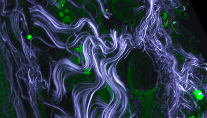
Three-dimensional rendering of mouse skin by Troy McClure. Autofluorescence caused by two-photon excitation in the green channel (pseudocolored green) and collagen SHG signal (pseudocolored grey).
When you think of Google Maps, what images come to mind? Satellite scans? Streetview photos? Graphical roadmaps? Users can zoom across insane scales of magnitude, understand how objects are related in space, and decide how to navigate—all in a click.
At the University Imaging Centers (UIC), mind-blowing new scopes are enabling biologists to begin constructing visual “maps” of life. The goal is to zoom seamlessly from the naked-eye view to the utterly small—but life is more complex than geography. Is it possible to visualize the tiniest phenomena at the fastest speeds and the deepest depths…all with one microscope?
Since the 1800s, optical microscopy had been stuck at a diffraction barrier of ~200 nanometers until a new family of Super Resolution scopes emerged several years ago. The UIC’s new Nikon Structured Illumination Microscope, a.k.a. SIMpson, magnifies down to a cool ~100 nm, and it’s wicked fast to boot. However, SIMpson (like Homer) is unfortunately a bit shallow.
On the other hand, the UIC’s new Nikon A1R MP multiphoton confocal, a.k.a. Troy McClure (yes, you guessed it—the UIC nicknames their scopes after all your favorite characters) can slice up to 2mm deep in a live specimen (like a mouse, pictured below) and blaze through over 400 images per second. Troy lacks SIMpson’s super-resolution, though you may remember it from such films as “Dial ‘M’ for Microscopy” or “Gone with the photon."
A single microscope can’t “do it all” due to different technical limitations in different situations, yet microscopy isn’t simply a matter of capturing pixels. In modern biology, microscopy is all about correlating big data across multiple image types and marrying that information to related findings from genomics, transcriptomics, proteomics and more. Microscopy helps us to see (and believe) what other biological fields hypothesize through more abstract evidence.
The UIC’s new instruments impart some powerful capabilities on the research community at the U, as do exciting new machines at the University of Minnesota Genomics Center and the Center for Mass Spectrometry and Proteomics. Stay tuned for more #CBSTech to learn about the most extraordinary machines on campus.
— Colleen Smith
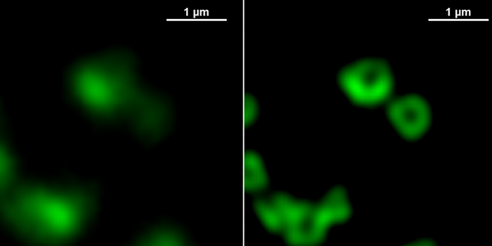
Synapses at the neuromuscular junction of fly larvae by SIMpson.

Troy McClure images neurons in a live mouse.

SIMpson's stage.

Troy McClure's eyepiece.