CBS researchers evaluate the efficacy of inhibition treatments in cancer cells

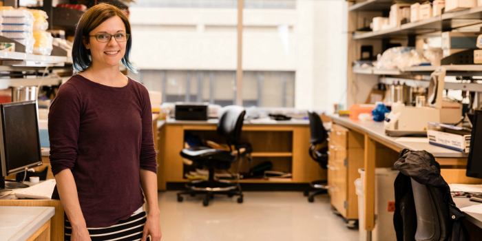
Professor Laurie Parker in her lab.
Like the many treats consumed at the State Fair, the enzymes within your cells can interact in unexpected ways. Until now, these interfaces have been difficult to detect. Laurie Parker in the Department of Biochemistry, Molecular Biology, and Biophysics, together with colleagues Wendy Gordon (BMBB), David Thomas (BMBB), and Paolo Provenzano (Biomedical Engineering) are pioneering a new technique to sort out how enzymes called kinases are reacting and interacting using fluorescence lifetime-imaging microscopy (FLIM). This research, funded by a grant from the National Institute of Health, seeks to map enzymatic activity in living cells and evaluate interactions between various enzymes to determine if complex relationships are disrupting the efficacy of current clinical treatments.
Gain of function mutations in kinases (which change their substrates to a phosphorylated state by adding a phosphate group) are frequently indicated in the development of certain cancers, and inhibiting their function has been an effective strategy for combating those diseases. But sometimes the inhibitors don’t work. One theory is that this is because of multifaceted exchanges within cells, wherein varying kinds of kinases are present, and the interactions between the many substrates and kinases decrease the usefulness of inhibition treatment.
Employing a microscope newly acquired by the University that has the capability to measure the spectral property of fluorescence lifetime, Parker and her lab have set out to find exactly where in the cell kinases actively function, individually and together. The Parker lab has developed peptide biosensors tagged with fluorophores that report kinase activity by producing different fluorescence lifetime states, depending on whether they are phosphorylated or not phosphorylated. Measuring the difference between the fluorescence lifetime decay rates of these two states allows them to calculate the kinase activity level within each individual cell. The fluorophores emit photons in response to excitation by light and the microscope can read these photons on a pixel by pixel basis to a very high degree of accuracy to calculate decay rates throughout the cell. The new microscope even allows for evaluation of two different combinations of probes at the same time, so multiple activities can be mapped together. Knowing this will help them to understand the sub-cellular location and overall level of kinase activity and, by proxy, what degrees of inhibition of the different target enzymes are being achieved in different cells. By elucidating the many interactions of kinases within living cells, these CBS researchers may be able to develop a method to use kinase activity as a surrogate biomarker for disease signaling in cancer patients. –Sarah Huebner