Post-doc fellow provides cinematic evidence that cells regulate tension forces during mitosis.
Think back to introductory bio—the cellular division unit. You probably got the big picture, but Jeremy Chacón, a post-doctoral fellow in the lab of Melissa Gardner (Genetics, Cell Biology and Development), unites concepts from biology and physics to measure the microscopic forces at play. He is lead author of a new study highlighted in the Journal of Cell Biology that provides a clearer picture of how it all works.
“The most vital task a cell needs to accomplish during mitosis is to put one copy of each chromosome into each daughter cell,” says Chacón. “If that doesn’t happen, cells can die, or worse, become cancerous.”
Scientists have long hypothesized that cells modulate tension to make sure chromosomes wind up in the right place, but they’ve lacked suitable measurement tools to rigorously test the theory. That’s where Chacón comes in.
At the Gardner Lab, Chacón studies how yeast cells sense and fine-tune the forces applied to migrating chromosomes. From a biophysical perspective, chromosomes behave like springs that obey Hooke’s Law—i.e. force equals stiffness times stretch.
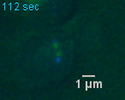
To calculate tension forces, Chacón first measures the intrinsic stiffness of chromosomes. Next, to measure stretch, he dyes chromosome pair centers (centromeres) green and the spindle poles located at the boundaries of each mitotic hemisphere blue. He uses high-powered microscopes to film metaphase in living yeast, and he writes software to analyze the videos.
Unlike real tug-of-war, the idea is to “tie” the game by lining up all the centromeres along the cell’s equator. This positions chromosome pairs properly so that eventually, when centromeres are cleaved in half, one chromosome goes to each daughter cell.
Chacón measured that microtubules accomplish this by applying a uniform force of 5 pN to each centromere. What’s even more amazing is the fact that when Chacón genetically modified chromosomes to become less stiff, cells responded by stretching centromeres even further, impressively maintaining a constant 5 pN force.
“No one’s been able to easily measure tension forces on this scale in live cells before,” says Chacón. “It’s been a big missing piece of the puzzle.”
— Colleen Smith